Technology

Ophthalmic Photography
Ophthalmic photography is a highly specialized form of medical imaging dedicated to the study and treatment of disorders of the eye. It covers a very broad scope of photographic services incorporating many aspects of commercial and medical photography. But it is through the use of highly specialized equipment used to document parts of the eye like the cornea, iris, and retina, that ophthalmic photography takes on it’s true identity.
The retina is the “film” of the eye. Images passing through the clear structures of the cornea and lens are focused there to give us our view of the world. Special instruments called fundus cameras, can show us the condition of this miraculous anatomical structure.
Optical Coherence Tomography
Optical Coherence Tomography (OCT)is a relatively and fundamentally new optical imaging technique developed for noninvasive cross-sectional imaging of the eye.
Breakthroughs in ophthalmic imaging make the retina’s inner workings transparent to a degree unimaginable even a few years ago.
OCT is a non-invasive technology used for imaging the retina, the multi-layered sensory tissue lining the back of the eye. OCT, the first instrument to allow doctors to see cross-sectional images of the retina, is revolutionizing the early detection and treatment of eye conditions such as macular holes, pre-retinal membranes, macular swelling and even optic nerve damage.
Similar to CT scans of internal organs, OCT uses the optical backscattering of light to rapidly scan the eye and describe a pixel representation of the anatomic layers within the retina. Each of these ten important layers can be differentiated and their thickness can be measured.
For certain conditions, such as age-related macular degeneration and cystoid macular edema, the OCT procedure is able to reduce or eliminate the need for fluorescein angiography for some patients.
Ultrasound
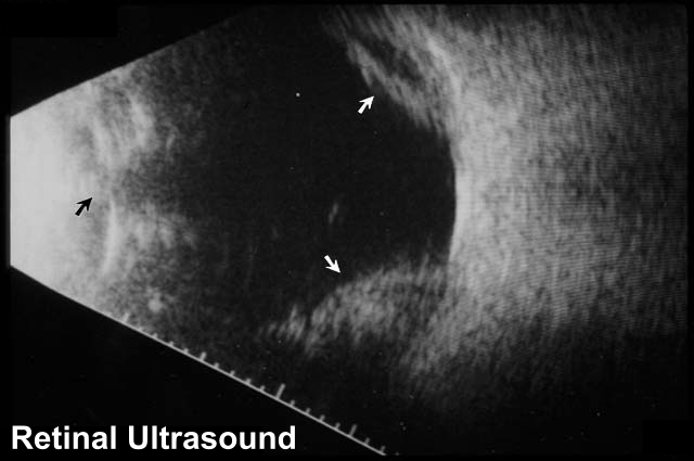
There may be occassions where your physician cannot view the retina due to some opacity that blocks the view – this is when we may use Ultrasound to determine the general status of the retina.
Ultrasound is a test that uses sound waves to assess ocular and retinal conditions and is commonly used to assess the retina in patients with a dense cataract or vitreous hemorrhage.
The best part for you? Ultrasound is simple to perform, painless, and does not involve any radiation.

Laser Treatment
A laser is a very focused beam of light that is used to treat a variety of eye disorders. The term LASER is actually an acronym coined from Light Amplification by Stimulated Emission of Radiation.
Laser treatment has revolutionized the treatment of a number of retinal disorders including diabetic retinopathy and retinal tears among others. For individuals with diabetes, laser treatment can significantly reduce the risk of visual loss both from leaking blood vessels causing retinal swelling, and from abnormal blood vessel growth (neovascularization). Finally, laser treatment around retinal tears significantly reduces the chance of a retinal detachment occurring.
Most commonly used lasers for retina procedures are the thermal lasers (argon and krypton). With these types of lasers, the light is converted to heat when it reaches the eye.
The heat is used to:
- seal blood vessels (veins and arteries) that are bleeding or leaking fluids;
- destroy abnormal tissue such as a tumor;
- bond the retina to the back of the eye
Laser treatment is almost always performed as an out-patient procedure. The patient sits comfortably on one side of the slit lamp (the microscope used to examine the eyes) while the treating physician is on the other side. Usually, a contact lens is placed on the eye to focus the laser after numbing eyedrops have been instilled. The treating physician then maneuvers a joystick which aims the laser in the desired location. The physician then steps on a footpedal or pushes a button to activate the laser. The laser burst lasts only 1/10th of a second and is usually accompanied by a clicking sound from the laser machinery. The individual being treated may see a bright flash of light.
Laser treatment of the macula (the central retina) is usually completely painless. Laser treatment to the peripheral retina usually requires a greater number of larger laser spots resulting in some discomfort (often described as a toothache-like pain). If a large amount of laser treatment is anticipated, the patient may opt to have the eye anesthetized by an injection of numbing medicine around the eye.
The results of laser treatment vary depending on the condition being treated and its severity. For many conditions, laser treatment is most effective at preventing visual loss and is best applied before visual loss has occurred (especially diabetic retinopathy).

